I spent a lot of time this semester familiarising myself with all the lab techniques involved in conducting an experiment in the Denny Lab. I was given a lot of responsibility, as well as independence, which meant a lot of the techniques were acquired on a steep learning curve. I am grateful to my supervisor for being so patient and encouraging throughout the training process.
New techniques:
Sectioning of brain tissues
In order to make uniform, thin sections the brain must be frozen. The apparatus we use is called a cryostat; it it maintains the platform-mounted brain and the knife inside a refrigerated compartment, to ensure that the brain remains frozen. I learned to first test if the brain was level, and to adjust the mount so that the sections would be even coronal slices, of 100 micrometer thickness.
Immunohistochemistry
This technique allows us to selectively image certain proteins, that act as antigens. Since antibodies specifically bind to antigens in biological tissues, primary and secondary antibodies are used to bind specifically to target antigens. This technique allows us to visualize and document specific cellular components, for example memory engrams, within their proper histological context. After the sections are stained, they are mounted and imaged. The process is time sensitive, as the sections have to be washed, blocked and exposed to the antibodies within a specific time frame.
Tissue Mounting
Mounting tissue is difficult because you have to mount the slices in order, i.e from anterior to posterior. This requires an intricate knowledge of mouse brain anatomy; luckily there is a mouse brain atlas at hand to guide me. This step precedes imaging and follows staining. After the brains have been mounted, we coverslip the slides so that they are ready to go for the microscope.
Confocal Microscopy
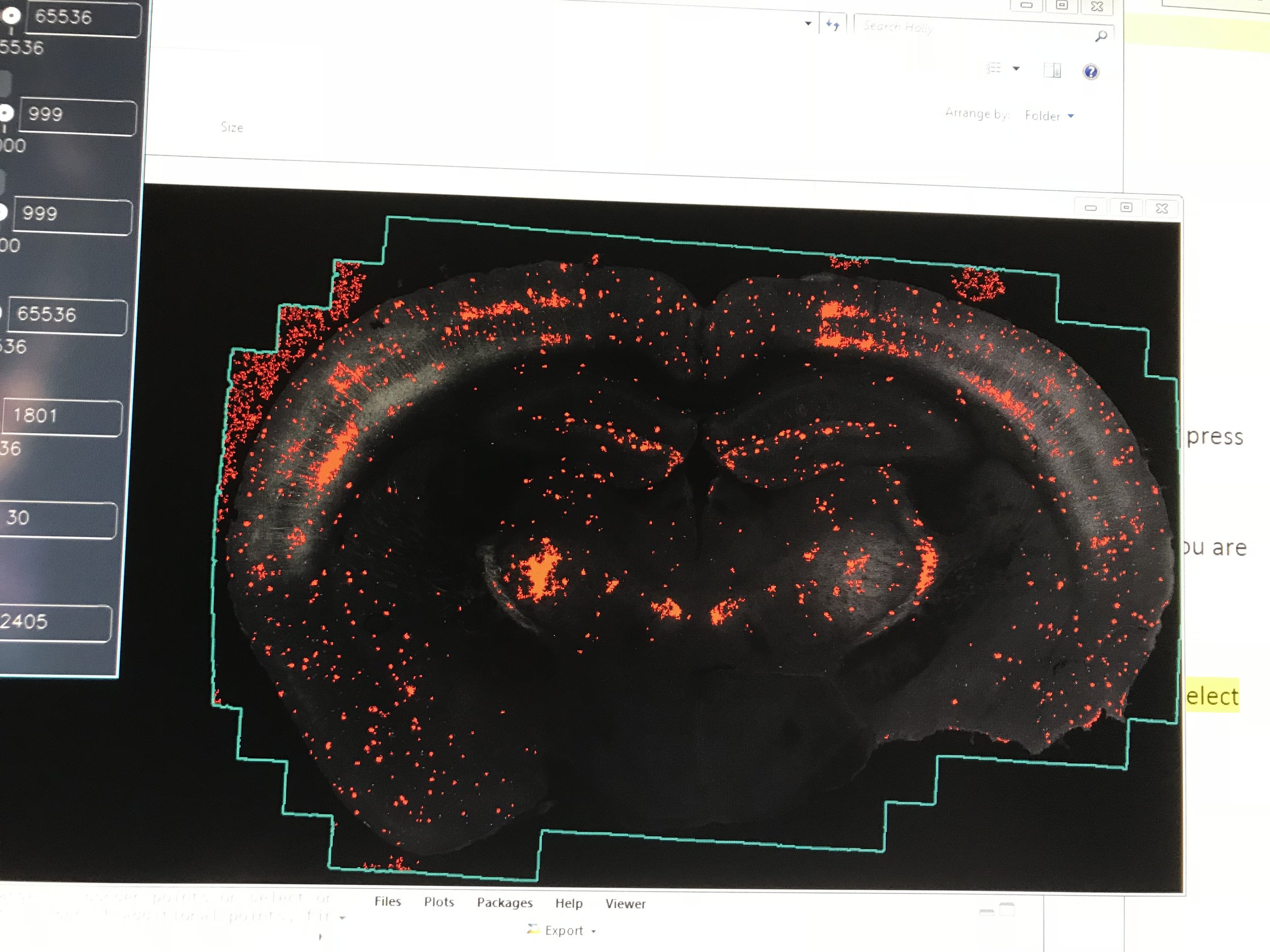
Fluorescent confocal micrographs were captured with a Leica TCS SP8. I was taught how to image the sections after mounting them on slides. The process consisted of a series of steps, and the software we use is pretty slow. After these images are obtained and converted, we cell count, which means counting the total numbers of, for example, EYFP and Arc cells in each confocal image and then counting the number of Arc cells that are co-labeled with EYFP. A ratio is then obtained, which tells us about memory creation or reactivation.
New knowledge:
Lateralization
In our third lab meeting if the semester, graduate student Jake Joran from another college presented his work on lateralization, specifically the potential functional lateralization in the hippocampus. We were all enthralled by his work and propositions.
Briefly, lateralization is the shift in specialised activity in one hemisphere. Studies have implicated that the left hippocampus, for example, is often associated with episodic, more externally driven memories. Whereas the right hippocampus is implicated to be associated with spatial, more internally driven memories. Jordan’s work seeks to investigate this further. I was inspired to pursue this phenomena in my research, potentially in relation to Alzheimer’s disease.
Impact of experimenter pheromones on rodent behavioural tests:
Behaviour testing is sex sensitive: when working with mice, one of the variables that has to be controlled is the sex of the experimenter. The pheromones of the experimenter will impact mouse behavioural tests. Since many projects in my lab monitor anxiety levels, what I learned is that the same sex has to, therefore, carry out all the tests. Apparently last year males and females alternated when handling the mice and this had a significant impact on the results. Good thing my postdoc both release the same pheromones!


